
Treatment of Laminitis

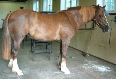
Laminitic foot – preparation for Imprint shoe
Objectives and observations of what may improve the patient’s recovery
By Andrew Poynton FWCF
The severity of a laminitic attack can vary so dramatically that horse owners respond in very different ways; some are quite unconcerned and assume it will all blow over and will all be ok soon, while others come out in a cold sweat and fear the worst.
Why might this be? Due to the many variables related to laminitis the outcomes vary widely; so, apart from any variation in treatment methods, the point at which remedial treatment commences also can sway the ultimate degree of recovery. Comparing one individual case of laminitis with another and what treatment may be the most effective is far from straightforward. Proponents of the various treatment methods often resort to anecdotal justification and pseudo science to bolster their particular preference.
Let’s take what often is the most straightforward case to treat, the small pony that is consuming more food than is good for it. It is likely to be carrying too much ‘condition’ (fat). Laminitic symptoms appear; the pony is lethargic, reluctant to move and stilted in its gait when persuaded to move. Providing remedial management is carried out in the form of reduced feed intake and an initial period of confinement, followed by gentle exercise at the appropriate time, all may be restored to harmony without farriery intervention.

Laminitic foot with the Imprint shoe grafted on
At the other end of the scale is the large horse with dramatic and severe onset of acute laminitis. This may have been triggered by one or more reasons not discussed here, but the attack is severe, the horse is heavy and when the lamellar bond is tentative, the weight of the horse tears this attachment asunder. Within hours, there is devastation within the hoof capsule. Within 24 to 48 hours a greater part of the union between the pedal bone and the hoof wall may already be seriously compromised (Pollitt 1995). Any attempt at remedial treatment following such a scenario is likely to be futile; total detachment of the pedal bone and descent within the hoof capsule from heel to heel is in my experience terminal. It is normally a question of time before the horse has to be destroyed, so regardless of treatment used, it is rarely effective. It must be appreciated that these are extreme ends of the scale, and this should be borne in mind.
Assessment and treatment
Before any additional materials are used in treatment of laminitics, the primary remedial work is to assess the hoof capsule in relation to the pedal bone, the relationship of the two, followed by expert remedial trimming as necessary. P3 is the point of reference for fitting the shoe, not the unstable hoof capsule.
It is essential that whatever is subsequently attached to the foot, if required, is precision fitted. The foot trimming is often 80 to 90% of the procedure. If this is carried out poorly or inaccurately, whatever products are used, the efficacy is going to be limited, if not detrimental. There is much talk about balancing horses’ feet, this is never more important than in an acute laminitic. The mediolateral and dorsopalmar balance are critical. Minimising leverage at point of breakover is advantageous.
Fitting of the toe of the shoe: It has been found to be helpful to fit the rim of the shoe in line as a continuation of the lower hoof wall or substitute, but in an improved position more closely aligned to P3 with the ground surface rounded to remove direct pressure or leverage on the dorsal wall. That way, sole clearance is maintained. By adopting this method the likelihood of the sole sinking onto the shoe is reduced and ample provision for shortened point of breakover is created.
How much extra length added to the hoof also has an effect on the laminal attachment? For the purposes of this discussion we will assume that the hoof capsule is trimmed to ideal proportions relative to P3. Once this is achieved, and as close to ideal digital alignment is attained, then support shoeing / treatment is carried out. This is where treatments begin to vary, as do individual theories of what is ideal and what is normal.
Perceived advantages of using thermoplastic shoes

P3 Imprint support
To begin with, early intervention is advocated and is possible due to the non-traumatic and efficient method of application. It can be applied without physical trauma to the foot, effectively and strongly so that the material forms as part of the hoof capsule. If fitted correctly, according to the instructions, it will be attached to the hoof until it is removed. Equally, it can be removed in a nontraumatic manner. It is radiographically translucent so X-rays can be taken while the shoes are in place. The material the shoe is constructed from is a low melt thermoplastic blend, which is completely malleable, allowing every part of this shoe can be adjusted. Therefore the attachment to the foot can be uniquely formed by softening the shoe material in hot water along with Imprint Hoof Repair material to rebalance the foot in any way desired and to form a frog cradle – a three-dimensional cast to the frog beneath the bony column, on which it can bear weight. This material does not crush under load, it holds its form so that when the potentially descending or rotating pedal bone pressurises the digital cushion beneath the pedal bone and the frog – both of these structures are compressible, but beneath that there will be the strong ‘frog cradle’, which provides support to the bony column; a finite base.

Before treatment
This limit for downward travel along with solar elevation, which is discussed later, can have a dramatic effect on the patient’s level of comfort. By providing this firm but accurate base, it is reasonable to assume there is reduced risk of mechanical dislocation of P3 and further lamellar tearing from the basement membrane.
That said, P3 is relatively small in comparison to the hoof capsule and contrary to common perception, when partially or greatly detached within the hoof capsule, it can move quite significantly, irrespective of what is attached to the outside. As we know in the healthy foot its lamellar attachment is designed to allow some degree of displacement as part of the suspensory and circulatory apparatus. Surrounded by venous plexus the colateral cartilages and digital cushion not to mention the deep flexor’s passage and navicular bursa, a somewhat different picture begins to emerge from that of the bone and horn of a freeze dried specimen.
After trimming & fitting Imprint shoes
It is for this reason that earliest possible application of the shoe is advocated. It is more prudent to apply support as a precaution rather than waiting to see what happens, especially in the larger equine. It is accepted that once lamellar breakdown has occurred, (Pollitt 1996) that union will not reattach but migrate away. If recovery follows then it is from new horn secreted from the papillae of the coronary corium. So early remedial attention could potentially limit the downward spiral of events, putting the brakes on the mechanical breakdown and begin to reverse the trend towards recovery from instability to stability.
It is worth noting that following radiographic diagnosis of some degree of rotational detachment of P3 and support shoeing has been undertaken, subsequent x-rays are likely to reveal further rotation, which often provokes alarm and negativity. This will be more evident in the larger equine. What seems often to be misunderstood is that the hoof capsule is rotating away from P3 much as a visor on a crash helmet is lifted or a fingernail lifting off following a traumatic blow. If there is heel growth also, which is often faster compared to any growth at the toe, this impression of impending doom is accelerated. This is the natural course of events, what varies is the speed at which this happens and how well the departure of the detaching hoof can be managed while the new hoof wall is growing down to replace it. Each time the foot is trimmed it usually necessitates heel trimming in the palmar third of the foot, and removal of the deviating lower dorsal wall while maintaining solar elevation.
If following accurate trimming and support treatment guided by radiographs an improved phalangeal alignment should be attained and confirmed by further X ray. Particular notice of the patient’s clinical condition before and after treatment should be noted, and the level of comfort should be maintained or improved. If there is deterioration, the procedure would need revisiting to remedy the situation. The most common reason for this is pressure on a sensitive area usually in the toe. Progress could be described as three steps forward and one step back. Any practitioner does well to correctly balance the foot and provide support beneath the centre of the bony column. It is worth defining what is meant by support in this text – contact with the structure without inducing loading onto it. By adopting this approach, the foot circulatory function can operate much as in the unshod foot. As the patient shifts their weight arterial supply can perfuse and venous return drains the digit.
When the shoe is fitted correctly, the point of breakover can be moved to suit the individual equine’s conformation and gait, not that it will be moving far while in the acute phase. The leverage at the toe can be reduced and moved and at any stage throughout the period that the shoe is in place. This can be adjusted by filing, rasping, cutting and modifying the shoe by adding or subtracting material. It is infinitely variable. The heels of the shoe can likewise be rasped down to counter the accelerated heel growth that causes P3 to gradually pitch forward between treatments. There is no limit on the adjustment factor to the shoe, and without trauma to the patient.
It is highly advantageous to maintain access to the sole of the patient’s foot for a number of reasons:
- There is access for treatment without removal of the shoe.
- The condition of the sole can be monitored. If the sole has perforated, it can be treated and dressed.
- Enclosing the sole creates a humid environment, which produces condensation, and this will induce softening of the horn structure of the sole, which is already compromised. (If the material is bonded to the sole then this problem can be avoided.)
- Possible increased risk of infection due to negative anaerobic bacteria present unless packed with anti bacterial dressing or direct bonding to the sole.
NB. The author does practise hoof casting and reconstruction in certain severe cases from which positive outcomes have been achieved, however, this is not discussed here.

Partial hoof slough a.
Keeping the sole uncovered if at all possible, dry and able to be disinfected on a regular basis has multiple advantages. Because of instability within the foot P3 bears down on the sole and its thin distal border induces pain pressing against the circumflex artery and solar venous plexus. Any upwards force or so called ‘support’ across the sole in this area can be counterproductive and induce further pain.
Where the sole can be elevated and unloaded from below, this usually achieves some relief for the patient. The bodyweight via the pedal bone, digital cushion and the frog are supported in a greater way in the acute laminitic because of the unnatural lamellar breakdown.
A main objective is to reduce stress on the compromised lamellar union. The shoe can be rounded on any face, rockered if required, but it is wise to maintain a level area of stability for the patient to rest upon. Additions and subtractions can be carried out during or after fitting as the condition progresses and time passes. It can be cut and rasped to counter any dislocation and growth of the hoof to keep the balance of the foot more ideal for a longer period. In fitting the shoe, minimal extra depth or weight is added to the hoof. The shoe virtually becomes part of the hoof. It can flex and move with the hoof. The central cleft in the frog plate also aids the heels to move three-dimensionally. Experience has shown that this mobility can greatly aid in pain relief in the acutely sensitive laminitic.

Partial hoof slough b.
In addition, it is helpful if the patient is bedded on a surface that is supportive but yielding on impact such as clean dry rubber flooring along with a moderate depth (6″ [15 cm]) of bedding covering the greater part of the floor. The author’s preference is large flake wood shavings or good wheat straw, as these are less likely to ball up in the foot and allow air to the sole. Fine beds such as sawdust or sand may be advocated by some as being supportive bedding, but in cases where sole pressure induces increased pain this bedding is less suitable. Given the option, the patient will quickly make it clear which surface is most comfortable as it walks on it.
A key objective when developing the system was to assimilate the structure and function of the foot as closely as possible with the materials used. This is why the plastics blend shore hardness is formulated to 65 D shore hardness, which is in the mid range of hoof wall hardness. This can be modified by the thickness of the plastic in any given area. Likewise, the frog plate central cleft is designed to allow normal heel flexion and hoof capsule movement. This can be adjusted by increasing the length of the cleft for more freedom of movement; conversely by reducing the length of the cleft by welding some plastic into it this will reduce mobility.
Each of these areas related to possible pain reduction can result in the accumulative effect of a significantly more relaxed patient. This will be evident in the patient’s demeanour – from a lethargic, depressed, stiff patient that is unwilling to move or even in a recumbent state where the musculature appears in spasm, to an interested, relaxed, mobile creature that is willing to pick up a foot and is interested in food. This frequently has the advantage of a reduced level of painrelieving medication needed to be administered. Having dealt with the source of pain via trimming and practical hoof support, if required any residual pain may be alleviated by medication. Reference to medication here is related to pain relief only and not to associated conditions such as equine metabolic syndrome (EMS) or Cushing’s disease.

Prosthetic hoof replacement.
Hoof sloughing can and does occur in more severe cases. Experience has shown that equines 14 hh upwards are more likely to succumb to this complication, due to carrying sufficient weight to more readily tear through the hoof. With such cases a second crisis period, post-acute stage, anywhere between two to four months into recovery is not uncommon. This is when the hoof capsule is sloughing but the new hoof has yet to regenerate and may be less than a quarter grown. The problem here is what is the horse going to bear weight on in the interim period?
It needs a replacement hoof for the time being while the new one grows. This is where the blend of low temperature thermo-plastics combined with specially selected adhesives come into their own. Following scrupulous preparation, a prosthetic hoof capsule can be sculpted onto the depleted hoof without risk of either chemical or heat burns. If applied intelligently, the material can closely assimilate the real thing. A shoe can then also be welded to the made up hoof at low temperature. This, can be very useful in assisting a patient through a tricky stage of recovery.
Complications
If complications are encountered, which inevitably will happen from time to time where a number of cases are treated, the most common will be infections around the distal border of P3. These can usually be accessed and drained via the hoof wall by cutting through the rim of the shoe and hoof wall without removing vulnerable sole.
If the sole prolapses and perforates, part of the ground bearing surface of the shoe can be cut away to allow access for treatment, while still keeping it elevated from the ground. Hospital plates can be quickly constructed from hoof repair material.
Finally, when considering what method of treatment to employ, it is worth first asking a few pertinent questions such as:
- What is the treatment designed to achieve?
- Does it work?
- Will it aid in P3 stability without compromising circulation?
- Can it be applied with minimal trauma to the patient?
- Is there likely to be an early positive response to treatment, eg, pain relief, ease of mobility, interest in food?
- Will any perceived benefits be maintained long term?
- Can the method and materials nurture the patient through all the stages of the condition to restored health?
- Is it worth it?
Reasoned thought and prior evidence may help in answering questions like these and ultimately provide more effective treatment of the laminitic equine.
References
Pollitt C.C. (1995) Development of laminitis. In Atlas of the Horse’s Foot. 171
Pollitt C.C. (1996) Basement membrane pathology: A feature of equine laminitis. Equine Veterinary Journal. 28. p146
Photographs: Andrew Poynton
From an article which first appeared in Forge magazine, February 2012. © Andrew Poynton FWCF
For advice or referral contact…
Imprint Equine Foot Care – Tel: +44 1666 822953
Poynton Farriery Clinic – Website: here